All of the power, none of the space
The power of an electron microscope needn't come at the cost of lab space says Mhairi Crawford of Hitachi High-Technologies
The power of an electron microscope needn't come at the cost of lab space says Mhairi Crawford of Hitachi High-Technologies
| |
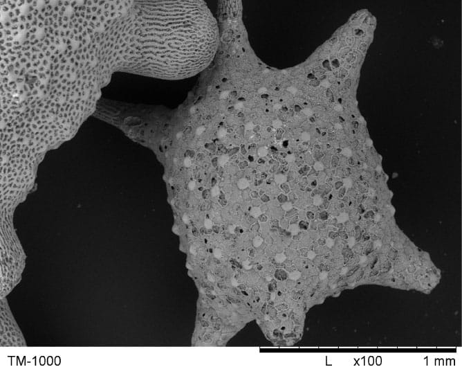
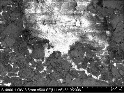
| Figure 1: Starsand |
This has been especially true in the world of microscopy, where electron microscopes have traditionally required a room to themselves, not just for reasons of their size but because of ancillary demands for pipes and services. The simpler optical microscopes, of course, can happily occupy the bench-top, but because their performance is limited by the wavelength of light, their powers of magnification and resolution are inferior to that of the electron microscope. Therefore, where magnification is required in excess of 1000x, or a resolution of much better than 0.3μm is needed, the optical microscope is unable to deliver.
Simple alternative 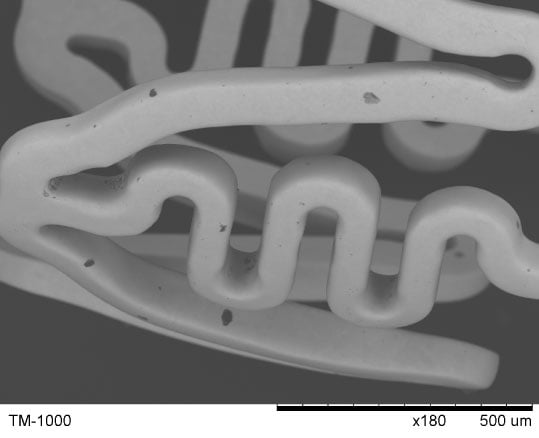
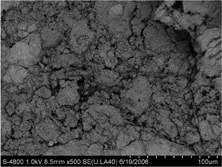
Figure 2: Stent, used to open arteries
In April 2006, at Analytica in Munich, the European laboratory market had its first sight of a new tabletop microscope from Hitachi-High Technologies, which bridges the gap between the worlds of optical and electron microscopes. Available in Japan since April 2005, the TM-1000 has already found applications in industrial, academic and government laboratories, examining material as diverse as cosmetics, stone, fabrics, electronic parts and pharmaceuticals.
With a footprint of just 564mm x 478mm, the TM-1000 will sit comfortably on a bench top but offers magnification and resolution performances much closer to that of a standard SEM. Indeed, the TM-1000 can be properly described as a scanning electron microscope, but it is greatly simplified for easy set-up and ease of use. In terms of set-up the TM-1000 is virtually ready for use as soon as it's delivered. No special facilities such as cooling water are required. It merely needs to be plugged in to a standard electrical supply. The 'Autostart' function turns on the beam, and automatically adjusts the magnification and focus, so that the specimen can be imaged immediately.
Whilst the tungsten filament used as the electron source is just the same as that used in a standard SEM, the accelerating voltage is set at a constant 15 keV. The rest of the instrument is optimised for this accelerating voltage, and so the TM-1000 is very stable.
The depth of field and resolution of images produced by the TM-1000 are superior to those offered by light microscopes as the high resolution image of star sand demonstrates (Figure 1). Moreover, it is capable of producing both topographical and elemental contrast detail. Figure 2 is that of a stent, used in medical applications for opening arteries, and the back scatter detector of the TM-1000 clearly exposes contamination on the surface.
Conducting samples, such as the PCB are readily observed because the entire chip can be put straight into the microscope with no sample preparation and the microscope itself is also easy to use. In fact, auto functions for focus, brightness and contrast, give the TM-1000 a 'point and click' functionality, such that anyone familiar with using a digital camera will be able to use the TM-1000 with very little training.
Non conductors
Non-conducting samples have always presented something of a challenge for the electron microscope, because the electron beam can cause charging on the surface of the specimen leading to damage and image distortion. The usual
 |  |
| Figure 4: Shirt during charge reduction mode | Figure 5: Shirt during high-vacuum mode |
Similarly, the image of pollen grains on a bee's-knee (Figure 6) clearly illustrates that the fine detail of biological specimens is maintained under this procedure and the resolution and depth of field again exceeds that of a light microscope. Very wet samples, such as cells and fully hydrated samples can also be imaged in the TM-1000.
High performance
As discussed above, the TM-1000 offers a 10x improvement in resolution and
 |
| Figure 6: This image is the Bee's knees. Literally. |
Samples of up to 70mm in diameter and 20mm thickness can be accommodated, and the TM-1000 features a magnification range of 20 – 10,000x using standard imaging and up to 40,000x using digital zoom capabilities. A built-in measurement function allows dimensional information to be acquired quickly and easily.
Revolution or evolution
The dedicated microscopy laboratory will, of course, always find a place for a conventional SEM. The TM-1000 was never intended to replace the conventional
 |
| Figure 7: White blood cells |
SEM, and experienced microscopists will continue to value the flexibility provided by a full range of accelerating voltages and detectors, and the higher resolution provided by such a system. Nonetheless, for routine observations, the TM-1000 could prove to be a useful addition to the laboratory. It will give microscopists a useful 'first-look' at samples, at the same time as freeing time on the conventional SEM for more detailed investigative work. Similarly, the general laboratory, which is perhaps frustrated by the limitations of their optical and stereoscopic microscopes, will undoubtedly find many reasons to welcome the greater magnification and resolution of this new SEM. And since it only occupies a modest space on the bench-top, they should also find many places where they can put it.
| Hydrated samples |
By Mhairi Crawford, Hitachi High-Technologies Corporation
Related Content

Siemens recognition for Bürkert enables improved development
Fluid control specialist Bürkert has become an official Siemens Product Partner for Siemen’s SIMATIC Automation Systems. The new partnership will …

Innovation in dialysis equipment
In 2022, more than 30,000 people were undergoing kidney dialysis in the UK [1], all of whom will require precision …

Liebherr launches new under-bench fridge and freezers for lab and healthcare providers
Liebherr, specialists in commercial refrigeration, have introduced its new range of under-bench fridges and freezers, engineered to meet the diverse …