
Flea bacteria

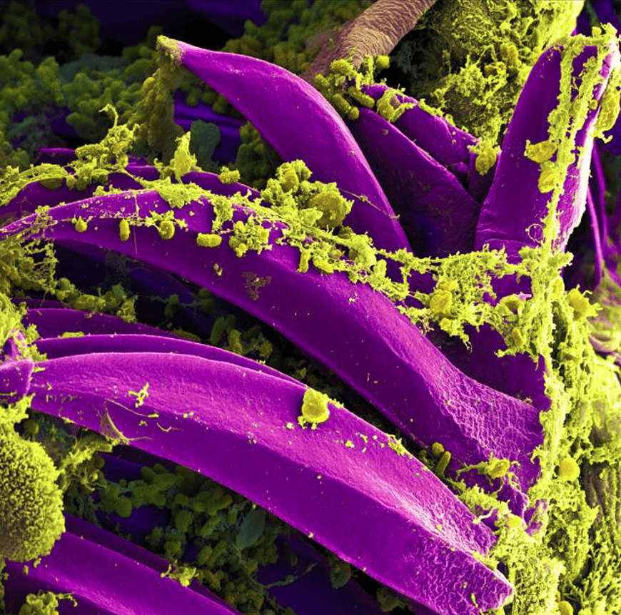
This digitally colourised scanning electron microscopic image depicts a number of yellow _Yersinia pestis_bacteria.
Produced by the US National Institute of Allergy and Infectious Diseases (NIAID), this digitally colourised scanning electron microscopic image depicts a number of yellow,_Yersinia pestis_bacteria, that had gathered on the proventricular spines of a_Xenopsylla cheopis_flea. The_Y. pestisbacterium is the pathogen that causes bubonic plague.
Related Content

Brain cells head to space in bid to advance neurodegenerative disease research
Researchers at the Saudi King Abdullah University of Science and Technology (KAUST) have sent two biomedical experiments to the International …

£3.75m programme targets rising UK mosquito and midge disease risks
A £3.75 million research programme led by the University of Glasgow will investigate how environmental change could alter the risk …

When seeing is no longer believing
Artificial Intelligence (AI) is polluting the world of science. Online teaching and reference resources are suddenly being populated by images …

