With alternative technologies available, we should be seeking antibody alternatives that avoid the unnecessary use of animals in research, urges Dr David Bunka.
Immunohistochemistry (IHC) is a vital technique in pathology and biomedical research that allows the visualisation and localisation of specific biomarkers in tissues. This powerful method is the gold standard test in clinical diagnostics for oncology and precision medicine, where it is used to identify tissue abnormalities, establish prognoses, and indicate therapeutic options. Researchers also use IHC to increase understanding of various molecular diseases and expand the range of novel disease biomarkers.
IHC has long relied on antibody tools as essential reagents that enable biomarker detection within relevant tissue sections. However, antibodies are associated with their own problems. Alternative technologies now offer the potential to overcome these problems and extend the scope of IHC with the potential for new biomarkers, improved performance and animal-free reagents.
Ethical compliance and reproducibility
Antibodies are animal-derived reagents generated by immunising animals with relevant targets. The resulting host animal’s immune response gives rise to the antibodies for use. Every year in the EU , approximately one million animals are used for antibody generation and production [1], despite the availability of technologies that do not necessitate the use of animals. Not only is this number high but the procedures employed often cause severe suffering.
As the advent of the FDA Modernisation Act 2.0 aims to move clinical development away from inaccurate animal models, there is potential to also move the production of research tools to more reliable and ethically compliant methods.
Beyond ethical concerns, the in vivo generation of antibodies can also lead to issues with lot-to-lot variability. Variability in polyclonal antibodies stems from differences in blood draws from the host animal. Within each draw the antibody mixture may vary each time and when the host animal dies the antibody becomes unavailable. Additionally, only between 0.5% to 5% of total antibodies in a polyclonal reagent are actually specific for the target [2].
Variability in monoclonal antibodies is due to hybridoma drift in cell lines producing the antibodies. This involves changes in the sequence of antibody-encoding genes over time as the hybridoma cells divide, which can lead to changes in the antibody and antibody lots over time. Both of these problems cause issues for scientists that need to be able to rely on their results and the reproducibility of their tools not just over the few years of a single research project, but over man y years to allow methods and results to be compared across projects.
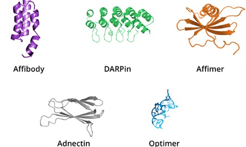
Newer antibody alternatives are presenting solutions to these problems. Binders, such as Adnectins, Affibodies, Affimers, DARPins and Optimers (Figure 1), are generated through in vitro discovery and development, removing the need for any animals in the process.
Once the binder is identified, they are manufactured through animal-free, controlled downstream processes that deliver improved lot consistency so scientists can rely on the reagent to provide the same result every time throughout the years of use without additional work to standardise lots.
Improving selectivity and sensitivity
A commonly cited problem with IHC antibodies is a lack of target selectivity. Researchers need antibodies to recognise the target antigen without cross-reacting with other proteins in the tissue sample. This seemingly simple requirement is often overlooked and has been repeatedly reported as causing issues in IHC analysis, even with validated antibodies used in pathology labs [3,4].
Antibodies typically recognise target epitopes consisting of only a few amino acids, which can be part of multiple proteins and peptides. For example, an anti–human proinsulin antibody cross-reacts with both insulin and glucagon-secreting cells. Because antibodies recognise a common epitope, the IHC reaction is virtually indistinguishable with cross-reactive proteins in the tissue from that with the intended target [5]. A specific advantage of nucleic acid and peptide aptamers, such as Optimer and Affimer, respectively, is their recognition of conformational epitopes [6,7]. The change in epitope recognition from linear to conformational of these antibody alternatives means that they can offer increased selectivity in performance. Additionally, the ability to include cross-reactive targets in the development process for these antibody alternatives ensures that any cross-reactive binders are removed during development.
A further challenge that can cause selectivity issues is the alteration of many antigens during the tissue fixation process. Tissue cross-linking with aldehyde fixatives can change the epitope structure, resulting in false positive results through antibodies crossreacting with non-target proteins or a false negative result in IHC as the antibody is unable to bind the target protein in the tissue. Of the newer antibody alternatives, Optimer development in particular can be performed using fixed samples.
This accounts for any changes in antigen structure that result from fixation and ensures the developed antibody alternative binds selectively to the required target in the format presented in the final IHC application for the best results in the final IHC assays.
Compared to standard IgG antibodies with a molecular weight of 150 kDa, most antibody alternatives have a molecular weight of 10-20 kDa. This smaller size offers advantages in IHC, as they can fit into smaller crevices on a target protein for more selective epitopes and to diffuse faster through tissue sections to speed staining. One study showed that for IHC application, Affimer ligands to VEGFR2, a key regulator in vasculogenesis, angiogenesis and associated with tumour neovascularisation, showed identical staining patterns to a control antibody. However, experiments showed that the Affimer staining developed faster than for the antibody, indicating a higher staining sensitivity [8].
Automated IHC
Automated IHC platforms, multiplex methods and digital imaging have contributed to decreased turnaround times and streamlined procedures. This has increased the effectiveness and precision of IHC to make it more affordable and practical for a range of clinical and research applications. To meet this growing market, antibody alternatives must be flexible enough to be incorporated into these automated workflows. However, as a result of over 80 years of antibodies being the mainstay as IHC tools, automated IHC processes are mainly based on antibody use.
While there are many arguments to embrace antibody alternatives, the transition to these newer tools will take time and the ability of these antibody alternatives to integrate into current platforms is essential. The new Optimer-Fc from Aptamer Group is an antibody alternative for use in automated IHC workflows, where it replaces the primary detection antibody in the assay (Figure 2) [9].
Capitalising on the highly selective and tuneable nature of Optimer binders, this development enables life scientists to pursue novel and existing disease biomarkers and offer increased accuracy and selectivity in IHC for research and diagnostics.
Animal-free antibody alternatives are now established reagents and offer many additional benefits for researchers from higher sensitivity, faster methods and lower costs as tools for IHC to the ability to integrate into current automated workflows for ease of adoption. With the move across the R&D pipeline to reduce animal usage, it is now important that we also embrace nextgeneration affinity ligands to support better research with antibody alternatives.
- Dr David Bunka is Chief Scientific Officer at Aptamer Group
References:
- Gray, A. C. Animal-derived-antibody generation faces strict reform in accordance with European Union policy on animal use. Nat Methods. 2020. 17, 755-756
- Bradbury, A. & Plu?ckthun, A. Reproducibility: Standardize antibodies used in research. Nature. 2015.518(7537):27-29
- Pisapia, D. J. & Lavi, E. VZV, temporal arteritis, and clinical practice: False positive immunohistochemical detection due to antibody cross-reactivity. Exp & Mol Path. 2016. 100(1) 114-115
- Lenggenhager, D et al. Immunohistochemistry for hepatitis E virus capsid protein cross-reacts with cytomegalovirus-infected cells: a potential diagnostic pitfall. Histopathology. 2023. 82(2):354-358
- Ramos-Vara, J.A. & Miller, M.A. When Tissue Antigens and Antibodies Get Along: Revisiting the Technical Aspects of Immunohistochemistry – The Red, Brown, and Blue Technique. Vet Path. 2013. 51(1): 42-87
- Davis, J.J. et al. Peptide Aptamers in Label-Free Protein Detection: 2. Chemical Optimization and Detection of Distinct Protein Isoforms. Anal. Chem. 2009. 81(9):3314-3320
- Zichel, R. et al. Aptamers as a Sensitive Tool to Detect Subtle Modifications in Therapeutic Proteins. PLOS One. 2012. 7(2):e31948
- Tiede, C. et al. Affimer proteins are versatile and renewable affinity reagents. eLife. 2017. 6:e24903
- LabMate Online. New reagent solution for immunohistochemistry. Accessed at: https://www.labmate-online.com/news/laboratoryproducts/3/aptamer-group/new-reagent-solution-forimmunohistochemistry/60232

